
Echocardiography (2D Echo) in Dwarka
Dr. Amit Pendharkar Senior Director & Unit Head – Interventional Cardiology
An echocardiogram, or echo, is an ultrasound scan of the heart. It shows how large the heart is, how well it pumps, and whether the valves open and close properly. The test is painless, uses no radiation, and takes about half an hour.
Dr. Amit Pendharkar, interventional cardiologist at Max Super Speciality Hospital, Dwarka, has performed more than 10,000 echocardiograms and stress tests, providing precise diagnostic evaluations for patients from Dwarka, Janakpuri, and West Delhi.
What Is an Echocardiogram?
An echocardiogram uses high-frequency sound waves to create moving pictures of the heart on a screen. A small probe covered in gel is placed on the chest and moved to different positions. Colour Doppler, which is added to a standard 2D echo, shows the direction and speed of blood flow, so leaking or narrowed valves can be accurately assessed.
- 100% painless, safe ultrasound imaging with zero radiation
- Precise evaluation of chamber sizes and ejection fraction (pumping power)
- Colour Doppler assessment for leaky or narrowed heart valves
- Direct review and report explanation by an experienced cardiologist
When Is an Echocardiogram Advised?
A cardiac ultrasound provides real-time moving images to identify structural changes, valve abnormalities, and pumping weaknesses before complications develop.
Breathlessness, chest pain, palpitations or swelling of the feet
A heart murmur heard by a doctor during routine check-ups
After a heart attack, to see how much the heart muscle has been affected
Long-standing high blood pressure or diabetes
Follow-up of known valve disease or heart failure
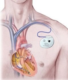
Before and after heart procedures such as angioplasty, TAVI or pacemaker implantation
Before major non-cardiac surgery in people with cardiac risk
What an Echo Can Show
| The echo checks | Why it matters |
|---|---|
| Size of the heart chambers | An enlarged chamber can point to long-standing high blood pressure, valve disease or heart failure |
| Pumping strength (ejection fraction) | Shows how well the heart squeezes and helps guide treatment |
| Heart valves | Detects narrowing (stenosis) or leaking (regurgitation) |
| Heart muscle movement | Areas that move poorly may have had a poor blood supply, for example after a heart attack |
| Fluid around the heart | Identifies pericardial effusion |
| Pressure in the lung circulation | Provides an accurate estimate of pulmonary artery pressure |
| Structural problems | Identifies holes in the heart (such as ASD/VSD) and other defects present from birth |
Why Choose Dr. Amit Pendharkar for Echocardiography in Dwarka
-
More than 10,000 echocardiograms and stress tests performed
-
A cardiologist reads the pictures and explains the result to you directly
-
DM in Cardiology from AIIMS, New Delhi, with more than 15 years of experience
-
If treatment is needed, angiography, angioplasty and other procedures are available with the same doctor at Max Super Speciality Hospital, Dwarka
-
Clinics in Dwarka Sector 6 and Janakpuri for consultation and follow-up
How the Test Is Done
No fasting is needed for a standard 2D echo. Take regular medicines unless told otherwise.
You remove clothing from the upper body and lie on your left side on the examination couch.
ECG stickers are placed on the chest to record the heart rhythm continuously during the scan.
Gel is applied and the ultrasound probe is moved over the chest while live pictures are recorded.
You may be asked to hold your breath briefly. The scan takes about 20 to 30 minutes.
You can resume normal activity straight away. The cardiologist reviews the pictures and explains the result.
Understanding Your Echo Report
The report lists chamber dimensions and a diagnostic summary. The most quoted number is the ejection fraction (EF). The table below provides a general guide; your cardiologist reads the whole report alongside your symptoms.
| Ejection fraction | General meaning |
|---|---|
| About 55% or more | Normal pumping strength |
| About 41% to 54% | Mildly reduced |
| 40% or below | Reduced; may indicate heart failure or heart muscle damage |
Types of Echocardiography
| Test | What it is | Used for |
|---|---|---|
| Transthoracic echo (2D echo with Doppler) | Standard scan through the chest wall | Most heart assessments and follow-up |
| Stress echo (dobutamine) | Echo pictures taken at rest and while the heart is stimulated with a medicine | Checking for blocked arteries in people who cannot do a treadmill test |
Dr. Pendharkar decides which echocardiographic method is suitable based on clinical evaluation and patient mobility.
Echo vs ECG vs Stress Test
| Test | What it records | Best at showing |
|---|---|---|
| ECG | The heart's electrical activity | Rhythm problems, signs of a past or ongoing heart attack |
| Echocardiogram | Ultrasound pictures of the heart | Structure, valves and pumping strength |
| Treadmill (TMT) / stress test | The heart's response to exertion | Blockages that appear only during effort |
While an ECG shows electrical signals, an echo displays mechanical function and physical anatomy directly.
Echocardiography for Patients in Janakpuri and West Delhi
Patients from Janakpuri, Vikaspuri, Tilak Nagar, Uttam Nagar, Palam and Najafgarh can see Dr. Pendharkar at his Janakpuri clinic on Mondays, Wednesdays and Fridays from 6 pm to 8 pm. Existing echo and ECG reports can be reviewed there, and any new scan is arranged at Max Super Speciality Hospital, Dwarka.
Frequently Asked Questions
Book an Echocardiogram Consultation in Dwarka
Call +91-9811675937 to book a consultation with Dr. Amit Pendharkar at Max Super Speciality Hospital, Dwarka, or at the Janakpuri clinic (Monday, Wednesday, Friday, 6 pm to 8 pm).